Women usually make their first visit to the gynecologist when they miss a period and suspect they are pregnant. Most of us have already taken a home pregnancy test.
At the first examination, which is usually performed up to the 12th week of pregnancy, the gynecologist reviews documentation and takes a medical history (personal and family). The midwife (or nurse) measures your blood pressure and body weight. You will receive a referral for blood tests (complete blood count, blood type, Rh-D factor, indirect Coombs test, first toxoplasmosis screening, blood sugar) and urine tests, which are often done at the gynecologist’s office (albumin, sediment, ketones, and glucose). The first ultrasound examination is performed, which is extremely important to determine the viability of the pregnancy, whether the pregnancy is intrauterine, and if heartbeats are present. The estimated due date is also determined. The gynecologist should discuss a healthy lifestyle and nutrition with you, and you will receive a maternity booklet that you must carry with you from the first day until childbirth.
However, problems requiring medical attention can arise in the first trimester.
- Bleeding in the first half of pregnancy – all bleeding is a pathological phenomenon. The main causes are spontaneous abortion, ectopic pregnancy, molar pregnancy, implantation bleeding, changes in the cervix, vagina, or external genitalia. In the case of a twin pregnancy, the death of one fetus occasionally occurs, which can lead to minimal uterine bleeding (Rakić, 2016).
- Spontaneous abortion – is the spontaneous termination of pregnancy before the fetus is viable – capable of survival in the external environment. It is the most common complication in the first trimester of pregnancy. It is estimated that approximately 15% of clinically recognized spontaneous pregnancies end in spontaneous abortion.
Fetal causes: chromosomal abnormalities in the fetus, congenital fetal anomalies, fetal injury (fetal injury due to necessary invasive procedures such as amniocentesis, chorionic villus sampling, cordocentesis).
Maternal causes: uterine abnormalities, microorganisms (Toxoplasma gondii, parvovirus B19, Listeria monocytogenes, measles virus, herpes simplex virus, cytomegalovirus, and lymphocytic choriomeningitis virus). And some other maternal diseases (thyroid dysfunction, adrenal glands, polycystic ovary syndrome, thrombophilia, autoimmune diseases).
And then idiopathic spontaneous abortions, which are abortions without a clear cause.
Clinical picture of spontaneous abortion is very non-specific. Vaginal bleeding occurs in 20–40% of women in early pregnancy. In the group of women where vaginal bleeding occurs between the 7th and 11th week of clinically confirmed pregnancy, spontaneous abortion does not occur in 90–96% of cases. On the other hand, 13% of women who miscarried did not experience vaginal bleeding. Spontaneous abortion is typically accompanied by cramping pain, similar to menstrual cramps (Došen, 2016).
Therefore, seek medical attention for any bleeding and cramping pain.
- Inflammation of female genitalia and pelvic inflammatory disease. In the reproductive age, numerous bacteria are present in the female vagina, forming the normal vaginal microflora. The normal vaginal pH is acidic, between 4 and 4.5. This acidic environment is maintained by lactobacilli, which produce lactic acid. The acidic environment of the vagina and the epithelium, which is rich in glycogen under the influence of estrogens, protect the vagina from infections. If the normal microflora is disrupted, which can happen, for example, after antibiotic treatment, after menstruation, after sexual intercourse, or due to systemic illness, other microorganisms can proliferate in the vagina, causing inflammation. A large proportion of infections are caused by sexually transmitted pathogens.
Vaginal and external genital inflammation – abnormal vaginal discharge (which may also smell), burning or itching sensation in the genitals. The most common are vaginal infections (colpitis, vaginitis), external genital infections (vulvitis), or both (vulvovaginitis). The most common causative agents are bacteria, followed by fungi and protozoa.
Bacterial vaginosis – burning and irritation sensation in the genitals, itching, grayish-white homogeneous and foul-smelling vaginal discharge. The discharge has a fishy odor.
Fungal genital infection – burning sensation in the genitals, itching, vaginal pain during sexual intercourse, genital erythema, genital edema. Characteristic is a white or yellowish-green vaginal discharge that adheres to the vaginal walls and has the appearance of cottage cheese or fresh cheese.
Genital infection with Trichomonas vaginalis – abundant, frothy, yellow-green, foul-smelling vaginal discharge, pain in the vagina and perineum, vaginal pain during sexual intercourse, painful urination, the external genitalia and vagina may be red, swollen, and pinpoint bleeding, resembling a strawberry surface, may appear on the vaginal mucosa and especially on the cervix.
In addition to Trichomonas vaginalis infection, other bacterial and viral sexually transmitted infections also clinically manifest as inflammation of the female genitalia.
Genital herpes – is also dangerous for the fetus and newborn. If a woman contracts genital herpes for the first time in early pregnancy, the chance of transmission to the fetus is small, but if it occurs, it can cause miscarriage. Pregnant women can also have a more severe form of the disease. If the first infection occurs towards the end of pregnancy, there is a high probability of transmission to the fetus.
Syphilis – Transplacental transmission of infection is most likely if the pregnant woman has early syphilis. In late latent syphilis, the probability of transmission is low. Fetal infection usually occurs between the 14th and 16th week of pregnancy. Syphilis infection can cause fetal death, fetal growth restriction, rhinitis, anemia, and other complications (Jančar, 2016).
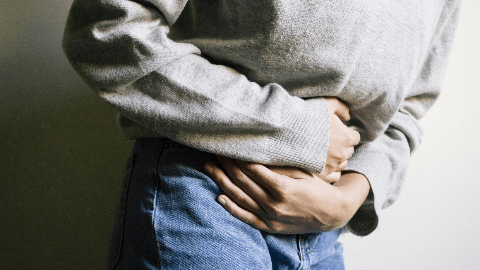
- Pain in the lesser pelvis (pelvic pain) is pain in the lesser pelvis and lower abdomen that can also radiate to the lower back and upper thighs. It can occur due to acute causes, meaning the pain lasts less than three months, e.g., due to ectopic pregnancy and urinary tract infection (uroinfection). Chronic – non-cyclic causes include, for example, constipation (obstipation), adhesions from previous surgeries, and others. Chronic pain lasts at least 6 months.
Ectopic pregnancy is located in the fallopian tube in 95% of cases. After approximately 4 weeks of pregnancy, it causes distension (distension) of the fallopian tube. This is a unilateral pain in the lesser pelvis, initially dull and of lesser intensity. The clinical picture then depends on the course of the disease. With spontaneous resorption of an ectopic pregnancy, the problems gradually subside. Most often, however, the contraction of the fallopian tube partially or completely expels the pregnancy into the abdominal cavity, leading to a tubal abortion with bleeding from the implantation site. In this case, the pain initially becomes cramping, then temporarily subsides, and a dull pain reappears, which can also spread to the entire abdomen, indicating dangerous bleeding into the abdominal cavity (intra-abdominal hemorrhage). In case of fallopian tube rupture, increasing dull pain is followed by slight relief and then a worsening of signs of bleeding into the abdominal cavity. In addition to pain, a characteristic 6–8 week missed period and less pronounced acyclic bleeding are also present. Therapy is mainly surgical with removal of the affected fallopian tube, less often medical treatment.
Torsion of uterine adnexa – torsion of the ovaries, fallopian tubes, or entire uterine adnexa causes these structures to twist around their axis. The most common symptom is unilateral lower abdominal pain, which is more frequent on the right side. However, the characteristics of the pain can vary. Problems begin suddenly after certain activities, such as lifting, physical exercise, sexual intercourse, turning in sleep, etc. The pain is often accompanied by nausea, vomiting, and loss of appetite (Reljič, 2016).
Urinary tract infection – microorganisms most commonly enter the urinary tract from the perineal area (perineum) via the urethra (ascending pathway). Due to the relatively short urethra, women suffer from urinary tract infections 30 times more often than men. Pregnant women have the same likelihood of urinary tract infection as the rest of the female population, but infections in them are more complicated and, in extreme cases, can be dangerous for the fetus itself, so urine in pregnant women must always be sterilized (even isolated bacteriuria must be treated). Thus, repeated urine tests are part of the standard monitoring or examination of a pregnant woman.
Urethritis – inflammation of the urethra is usually a consequence of infection with sexually transmitted microorganisms (especially Chlamydia and Gonococcus). The infection can be asymptomatic or manifest with burning pain in the urethra and/or discharge.
Cystitis – inflammation of the bladder usually manifests with frequent and burning urination, constant urge to urinate, and less often, bloody urine. Patients often also report pain above the bladder or above the pubic bone (suprapubic pain). Body temperature is usually not elevated.
Pyelonephritis – in inflammation of the kidney and renal pelvis, fever (38°C and above) and chills appear, lumbar pain, usually also problems due to associated bladder inflammation (Uroweb, n.d.).
Sources:
- Došen, M. (2016). Gynecology and Perinatology. In I. Takač and K. Geršak (eds.), Spontaneous Abortion (pp. 120-125). Maribor: Faculty of Medicine.
- Jančar, N. (2016). Gynecology and Perinatology. In I. Takač and K. Geršak (eds.), Inflammation of Female Genitalia and Pelvic Inflammatory Disease (pp. 130-140). Maribor: Faculty of Medicine.
- Rakić, S. [Saša]. (2016). Gynecology and Perinatology. In I. Takač and K. Geršak (eds.), Bleeding in the First Half of Pregnancy (pp. 484–487). Maribor: Faculty of Medicine.
- Reljič, M. (2016). Gynecology and Perinatology. In I. Takač and K. Geršak (eds.), Pelvic Pain (pp. 113–119). Maribor: Faculty of Medicine.
- Uroweb. (n.d.). Retrieved from Link
Photo: Canva