The breast lies between the fourth and seventh ribs. It is bordered by the sternum on the medial side and the midaxillary line laterally. More precisely, it lies on the fascia of the pectoralis major muscle and also extends to the lateral part of the chest wall (Arko, 2006). The breast consists of glandular, fatty (Geddes, 2007), and connective tissue (Arko, 2006). As Inch (2014) notes, the presence and amount of fatty tissue varies from woman to woman. This fatty and glandular tissue is supported by a loose layer of connective fibres, also called Cooper’s ligaments. Their role is to support the breast by connecting the deep fascia of the pectoral muscle with the superficial fascia. The breast is divided into 15–20 separate lobes (Geddes, 2007). These are glandular units, and each has its own duct that opens at the nipple (Arko, 2006). Each lobe contains lobules, which are made up of alveoli and ducts (Inch, 2014). They are separated by septa of connective and fatty tissue (Arko, 2006). As Geddes (2007) states, each lobule contains 10 to 100 alveoli. These are surrounded by capillaries (Kent, 2007).
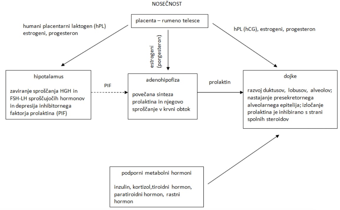
During pregnancy, the breast goes through certain developmental processes. This is a complex sequence of events controlled by hormones. At the beginning of pregnancy, the development of ducts, branching, and the formation of lobules can be observed. All of this is triggered by luteal and placental hormones. Placental lactogen, prolactin, and chorionic gonadotropin also contribute to accelerated breast development. Prolactin is essential for the full development of lobules and alveoli. This hormone, along with other protein hormones found on the surface of alveolar cells, controls its activity through receptors and influences the onset of milk release. To induce milk synthesis, cell division must be induced with the help of insulin and cortisol. Figure 1 shows that prolactin is secreted by the pituitary gland and is controlled via a negative feedback loop by prolactin inhibitory factor (PIF), which is secreted by the hypothalamus. The amount of catecholamines affects the release of PIF from the hypothalamus into the bloodstream. In the third month of gestation, the appearance of a colostrum-like substance can be observed. Prolactin stimulates colostrum production. By the second trimester, it may begin to be secreted, which is also due to prolactin. A woman who gives birth after the 16th week of gestation will secrete colostrum. During pregnancy, estrogen is thought to increase prolactin production, and this relationship is thought to be partly controlled by placental lactogen, which interferes and inhibits prolactin production. However, the main inhibitor of prolactin during pregnancy is progesterone. Estrogen also stimulates the stretching of the epithelial cells of the ducts. Although estrogen appears to promote breast development both directly and indirectly, it does not play an indispensable role in the development of this organ during pregnancy (Lawrence and Lawrence, 2011b).
Insulin and a growth factor normally found in human serum stimulate the proliferation of mammary stem cells. These cells later develop into alveolar cells under the influence of corticosteroid hormones. High prolactin levels during pregnancy are not a result of milk synthesis; rather, they are partly due to the effect of progesterone antagonism on prolactin’s stimulatory effect on casein in the synthesis of messenger ribonucleic acid (mRNA). In late pregnancy, lactogenic receptors, which have the same affinity for both prolactin and human placental lactogen, show a greater tendency to bind human placental lactogen. High doses of estrogen inhibit prolactin’s action on milk secretory cells. Likewise, elevated progesterone levels prevent its action. Abundant milk synthesis begins only after birth, when progesterone and estrogen levels drop. The key hormones that enable the onset of lactation are prolactin, insulin, and hydrocortisone (Lawrence and Lawrence, 2011b).

Figure 1: Preparing the breasts for lactation during pregnancy (Lawrence and Lawrence, 2011b)
Lactogenesis
Phase I, according to Lawrence and Lawrence (2011b), begins in the 12th week of gestation and is expected to end on days 3–4 after birth, while Riordan (2010) claims that this phase ends on day 2 after birth. In this phase, the mammary glands develop the ability to secrete milk components (Kent, 2007). It is marked by a sharp increase in lactose, total proteins, and immunoglobulins, and a decrease in sodium and chloride in the bloodstream (Lawrence and Lawrence, 2011b). As Kent (2007) notes, in late pregnancy a woman is expected to secrete about 30 ml of colostrum per day. Because it is not removed by the newborn’s suckling, the components are reabsorbed into the bloodstream. Lactose is not broken down in the blood and is therefore removed through the kidneys, i.e., in urine. The presence of lactose in urine therefore confirms the onset of lactogenesis I and, at the same time, pregnancy (Kent, 2007).
After birth, the breasts are still exposed to hormones that prepare them for breastfeeding, which is also shown in Figure 2 for easier understanding. A sudden drop in progesterone after birth triggers type II lactogenesis. Prolactin levels in plasma remain high. The onset of this process does not depend on the newborn’s suckling until the third or fourth day after birth, when milk secretion may stop if milk is not removed from the breasts (Lawrence and Lawrence, 2011b).
Soon after birth, type II lactogenesis begins. This is when a larger amount of milk starts to be produced (Kent, 2007). Riordan (2010) states that this phase begins on the third day after birth and lasts until the eighth day. Kent (2007) says its onset ranges between 24 and 102 hours after birth. In this phase, there is also a shift from endocrine control of milk synthesis to autocrine control. Prolactin secretion is still controlled by the hypothalamus (Riordan, 2010). This includes increased blood flow through the breasts and improved absorption of oxygen and glucose into the blood. A large rise in citrate concentration is also observed. All of these are thought to be indicators of the onset of phase II lactogenesis. Biochemically, its onset is signalled by an increase in the concentration of α-lactalbumin in plasma (Lawrence and Lawrence, 2011b). A prerequisite for its onset is a sufficient amount of prolactin, insulin, and cortisol (Kent, 2007). After the placenta is delivered, progesterone concentration in the blood drops, which stops its inhibitory effect on milk synthesis (Kent et al., 2010). In addition to the hormones mentioned, thyroid hormone is also important for the onset (Kent, 2007). As Riordan (2010) mentions, a drop in estrogen could also influence the onset of lactogenesis II. A woman experiences this phase as a sudden feeling of breast fullness. With each feed, oxytocin is released into the breastfeeding mother’s bloodstream. How much milk the baby gets depends on the number of milk ejection reflexes, not on how long the newborn is latched to the breast. With the onset of lactogenesis II, not only does the volume of milk change, but so does its composition (Kent, 2007). It continues to change until the tenth day after birth, when it transitions into so-called mature milk. The phase of mature milk secretion is called galactopoiesis, or lactogenesis III (Lawrence and Lawrence, 2011b). Galactopoiesis lasts until the onset of involution of the mammary glands. If a woman wants to breastfeed, regular milk removal is extremely important in this phase, as it stimulates the activity of the hypothalamus (Riordan, 2010).

Figure 2: Preparing the breasts for lactation after birth (Lawrence and Lawrence, 2011b)
Read also:Hormones important for breastfeeding
Source: Thesis “A woman’s experiences and feelings during breastfeeding” (2016) by Teja Šircelj, supervised by Senior Lecturer Tita Stanek Zidarič, Grad. Midwife, MSc, IBCLC