
Analgesia is defined as partial insensitivity to painful stimuli while consciousness is maintained (Močnik Rožnik, 2012). It is most commonly used during childbirth, as it helps the mother relieve labor pain while allowing the natural processes of labor to unfold smoothly. The mother simply loses the intense sensation of being able to feel pain in the lower part of her body. However, the sense of touch, pressure, and the ability to move remain unchanged (Simkin, 2012a).
Anesthesia is a blockade of all sensations, including pain. It is most often used for operative deliveries, as it also involves the loss of touch sensation, loss of certain reflexes, and loss of the ability to move in the lower body during labor. Some forms of anesthesia, such as general anesthesia, are used during labor and cause loss of consciousness. Other forms, such as regional anesthesia, remove all pain sensations from specific parts of the body while consciousness is maintained (Anesthesia, 2012).
January 19, 1847, is considered the beginning of obstetric anesthesia. James Young Simpson (professor at the University of Edinburgh) was the first to use diethyl ether for delivery in a woman with a deformed pelvis. Later, he also used chloroform for this purpose. On November 20 of the same year, he published his achievements in The Lancet. In 1853, John Snow used chloroform for anesthesia during Queen Victoria’s eighth delivery. Fifty years later, Steinbuchel used opiates in combination with scopolamine to induce amnesia and comfort during childbirth. In 1809, Serturner isolated codeine morphine from crude poppy seed extract, marking the beginning of percutaneous use. Klikgowich described the use of O (dinitrogen monoxide – laughing gas) in 1868, and Gusserow described acetylsalicylic acid for pain relief during childbirth (Brunček Gostenčnik and Wagner Kovačec, 2010).
Epidural analgesia was first used in 1898 for forceps delivery (Moir, 1986). Olah (1994) describes the usefulness and better accessibility of epidural analgesia only in the late 1970s. This was partly due to the decline in home births and partly due to increasing active intervention in the birth process, as added by Lieberman and O’Donoghue (2002).
Today, women have numerous methods available for labor pain relief, among which epidural analgesia and combined spinal-epidural analgesia are the most effective (Bofill et al., 1997; Sharma et al., 1997; Howell, 2000). However, epidural analgesia and combined spinal-epidural analgesia are not a complete guarantee of greater satisfaction with the birth experience (Zakšek and Grmek, 2011).
Epidural Analgesia
Epidural analgesia is an effective method for labor pain relief, but unfortunately, it begins to work with a 20 to 30-minute delay. Analgesia is achieved with a continuous infusion of a local anesthetic (in a dose of 10 ml of 0.2% ropivacaine solution, 0.125% bupivacaine solution, or chirocaine) or a combination of a local anesthetic and an opiate (in a dose of 10-12ml of 0.0625% bupivacaine or chirocaine + 2µg fentanyl/ml LA or 0.125% bupivacaine or chirocaine + 1µ fentanyl/ml LA). The infusion is started immediately after verifying the position of the epidural catheter and excluding its placement in a subdural or subarachnoid vessel. In cases of pain that increases, especially during the pushing phase, during operative vaginal delivery (“low” or “high forceps”), or during episiotomy repair, a bolus dose of 3-10 ml of 1.5% lidocaine solution or 0.5% ropivacaine solution is added (the desired block level is Th 10) (Stopar Pintarič, 2009).
EPIDURAL SPACE
The epidural space extends from the foramen magnum to the sacral hiatus (Brunček Gostenčnik and Wagner Kovačec, 2010). Nerve roots emerge from the spinal cord into the epidural space, innervating the uterus and birth canal, which is why epidural analgesia is most often injected into this space (Pang and O’Sullivan, 2008). It is bounded anteriorly by the posterior longitudinal ligaments, laterally by the pedicles and intervertebral foramen, and posteriorly by the ligamentum flavum. Within the epidural space are nerve roots, fat, lymphatic tissue, and blood vessels, with a well-organized Batson’s venous plexus. The epidural space is segmented and discontinuous. CT epidurography has revealed the presence of epidural septa. This can explain the occurrence of unilateral or incomplete epidural anesthesia. Ultrasound and magnetic resonance imaging have determined the distance between the skin and the epidural space, which measures 3-9 cm (average 4.5-5.5 cm). The structures traversed during catheter placement are: skin, subcutaneous fat, supraspinous ligament, interspinous ligament, and ligamentum flavum (Brunček Gostenčnik and Wagner Kovačec, 2010).
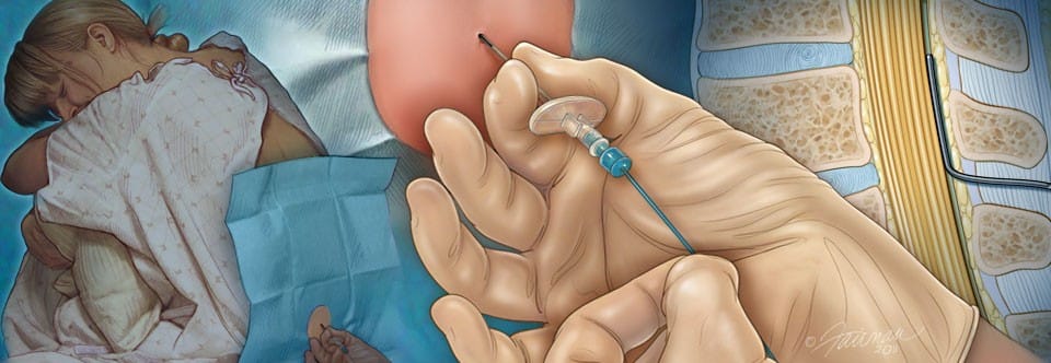
The doctor inserts the catheter into the epidural space using a special puncture needle with a diameter of 16 to 18 G. When pain needs to be relieved in the lower abdomen or lower limbs, it is inserted between the second and fourth lumbar vertebrae (L2 – L4); when pain needs to be relieved in the chest or upper abdomen, it is inserted between the fifth and eighth thoracic vertebrae (T5-T8) (Šmitek and Krist, 2008).
EA ADMINISTRATION
It is desirable that the mother is informed about the technique and method of the procedure. In some places, a prior anesthesiological examination in an anesthesiology clinic during late pregnancy is required, with prior submission of laboratory results (CBC, coagulation tests). For healthy mothers when the cervix is 4 cm dilated, the epidural catheter is placed in a lying lateral position or a sitting position, but earlier for high-risk mothers. Skin preparation for the puncture site must adhere to all surgical rules of antisepsis and sterility. In addition, an anesthesia machine with monitoring for a possible resuscitation procedure must be available. Epidural space puncture is performed in the midline, in the intercristal space at a distance – or – (the distance connecting both iliac crest bones). The needle used for the procedure is blunt with a side opening and a stylet (Tuohy needle), size 16-18 G. The puncture site is pre-anesthetized with a thin needle and a local anesthetic. The epidural space is identified using two methods: loss of resistance and the hanging drop technique. The loss of resistance occurs when the plunger of a filled syringe slowly advances when pressure is applied to the needle, which is connected to a saline syringe. The hanging drop phenomenon occurs when a drop located at the needle’s entrance is sucked into the epidural space due to negative pressure. To verify the epidural space, a catheter is inserted 4 cm into the epidural space (sensory block at the Th10 level). The catheter’s position is tested by applying a local anesthetic (e.g., 2 mL of 2% lidocaine with adrenaline) (Matošević et al., 2005).
INDICATIONS AND CONTRAINDICATIONS for Epidural Analgesia
Stopar Pintarič (2009) believes that the mother’s desire (in the absence of contraindications) is a sufficient indication for labor pain relief. Brunček Gostenčnik and Wagner Kovačec (2010) state that labor pain relief is particularly important for mothers where the body’s stress response to pain affects a fragile balance due to co-existing conditions. It is known that blood pressure fluctuations are smaller in eclamptic mothers, placental blood flow is improved, and regional anesthesia is safe to use, in case of the need for delivery by cesarean section. In mothers with diabetes, blood sugar fluctuations are smaller with effective pain relief. The hemodynamic effect is mitigated (sudden increase in preload, tachycardia, increased systemic vascular resistance, hypertension, hyperventilation) due to pain from contractions in mothers with other co-existing conditions (mitral stenosis, spinal cord injuries, intracranial vascular diseases, asthma) (Brunček Gostenčnik and Wagner Kovačec, 2010).
Indications also include multiple pregnancy and breech presentation, as well as post-cesarean section status. Epidural analgesia is also chosen in cases of failure to progress in labor or if instrumental delivery is required (May and Leighton, 2007).
Chestnut (2004) believes that every anesthesiologist must also consider the risks and benefits for each patient individually. Absolute contraindications include the mother’s refusal of epidural analgesia or her inability to cooperate (Sass, 2011). Pre-existing neurological and orthopedic conditions in the mother are also contraindications to epidural analgesia (Chestnut, 2004). Sass (2011) further adds severe coagulopathy, active infection at the catheter insertion site, and increased intracranial pressure. Epidural analgesia is also contraindicated if the team is not proficient in the technique of obstetric epidural analgesia, and if, due to insufficient knowledge or inadequate equipment, they are unable to manage complications (Zabavnik and Vajndal, 2001; Brunček Gostenčnik and Wagner Kovačec, 2010). Šmitek and Krist (2008) also list receiving anticoagulants, decreased level of consciousness, and hypovolemia in the mother among the contraindications.
POSSIBLE COMPLICATIONS
Risks during epidural analgesia use
If you have epidural labor analgesia, there is a 14% chance that instrumental assistance (vacuum, obstetric forceps) will be needed during the baby’s expulsion. Without EPA, this probability is 7% (translator’s note: In Slovenia, it is 3%).
With epidural labor analgesia, the second stage of labor (from full cervical dilation to birth) is usually longer, and there is a higher chance that you will need medication (oxytocin) to accelerate and strengthen contractions.
There is a higher chance of your blood pressure dropping.
During EPA, you may experience a feeling of weak legs.
You will have more difficulty urinating. You may need a catheter inserted into your bladder to drain urine.
Skin itching may occur.
An elevated body temperature may occur, which is not related to infection and may be accompanied by signs of fetal distress.
If you receive higher doses of opioids during EPA, it is possible that the newborn will need temporary breathing assistance, and the chance of successful breastfeeding may also decrease.
Other risks
On average, epidural labor analgesia does not increase the risk of headache. However, in approximately 1 out of 50 women who receive EPA, the epidural needle punctures the dura mater, which contains cerebrospinal fluid (this is called a “dural puncture”). If this happens, the likelihood of a severe headache, which can last for several days or weeks if untreated, significantly increases. If you experience a severe headache after birth with EPA, talk to your anesthesiologist, who will explain treatment options.
The following information is based on observational study findings
The risks of epidural and subarachnoid labor analgesia are shown in the table.
In approximately one out of 13,000 women, epidural labor analgesia causes long-term nerve damage. This results in problems such as muscle weakness or a tingling or numb sensation down one leg. However, nerve damage can occur both with and without EPA during childbirth. Nerve damage is approximately five times more common without the use of epidural labor analgesia, affecting one in 2,500 mothers.
There is no evidence that the use of epidural analgesia during labor causes permanent inflammation (i.e., swelling and tenderness) of the spinal nerves. If you are concerned about the risks of serious problems that may occur with EPA, discuss them with your anesthesiologist.

If you wish to relieve pain during labor with epidural analgesia, inquire at the maternity hospital where you plan to give birth whether this service is available, whether it is chargeable, and whether it is available 24/7. Also, find out if you need an additional consultation with an anesthesiologist, perhaps attending a lecture at a parenting school.
A leaflet is also available: Pain Relief During Childbirth by the Slovenian Association for Anesthesia and Intensive Medicine, from which I drew the possible complications used in this article.
Otherwise, the source of the article is the diploma thesis: “The Role of the Midwife in Epidural Birth (2012)”, by Sandra Topič under the mentorship of Senior Lecturer Teja Škodič Zakšek, B.Eng. Rad., B.Midw., MSc (UK).


