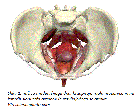
The pelvic floor divides the pelvic cavity and the perineum. It is composed of muscles and connective tissue. Two levator ani muscles attach peripherally to the pelvic wall and unite along the longitudinal axis with strong connective tissue. Together, they form the largest component of the bowl-shaped structure known as the pelvic diaphragm, which posteriorly terminates with the coccygeus muscle. This muscle extends over the sacral ligaments between the sacrum and coccyx to the ischial spine. The pelvic diaphragm forms most of the pelvic floor and has a deformed U-shape in its anterior parts, due to the urinary and reproductive organs located in the pelvic cavity. The anal canal passes from the pelvis to the perineum through a posterior circular opening in the pelvic diaphragm. The pelvic floor is anteriorly supported by the perineal membrane and the muscles beneath the membrane, the deep perineal muscles. The perineal membrane is a thin, triangular connective tissue that fills the space between the branches of the pubic arch and ends freely posteriorly towards the coccyx. The deep perineal muscles are a narrow area above the perineal membrane. The vagina and urethra penetrate the pelvic floor from the pelvic cavity to the perineum (Drake et al, 2005).

The pelvic floor muscles extend from the sacrum and coccyx to the ischium and pubis. These muscles support the pelvic cavity organs, move the sacrum and coccyx, and control the passage of excretions through the openings (Martini et al, 2006).
The pelvic floor is important for providing support to the pelvic organs and for maintaining continence during defecation and urination. It also plays a significant role in sexual intercourse. During pregnancy, the passive movement of the fetus through the birth canal affects the pelvic floor, causing it to relax and prepare for childbirth (Myles, 2009). The pelvic floor muscles extend from the sacrum and coccyx to the ischium and pubis. These muscles support the pelvic cavity organs, move the sacrum and coccyx, and control the passage of excretions through the openings (Martini et al, 2006).
The perineum is the inferior region of the pelvic floor between the lower limbs, extending across the entire pelvic outlet. It is diamond-shaped, with its inferior boundary being the pelvic outlet and its superior boundary being the pelvic diaphragm. It is defined anteriorly and superiorly by the symphysis, posteriorly by the coccyx, and laterally by the ischial tuberosities. The lateral boundaries are determined by the pubic arch anteriorly and the sacrotuberous ligament posteriorly. The main artery and nerves in this region are the pudendal artery and pudendal nerves (S2 to S4 roots). An imaginary line between the ischial tuberosities divides the perineum into two triangles. The anterior is the urogenital triangle, and the posterior is the anal triangle. It is important to note that these two triangles do not lie in the same plane. In the anatomical position, the urogenital triangle is oriented horizontally, while the anal triangle is directed downwards along the transtubercular line, facing more posteriorly. The “roof” of the perineum is largely bounded by the levator ani muscle, which divides the pelvic cavity above from the perineum below. These muscles are on each side, conical in shape, and terminate at the posterior tip of the anal triangle, forming the anal opening (Drake et al, 2005).
The superficial muscles of the urogenital triangle are the muscles of the external genitalia. These cover deeper muscles that reinforce the pelvic floor and surround the urethra. These muscles form the urogenital diaphragm, a deep muscular layer that extends between the pubic bones. Even more extensive muscular tissue, called the pelvic diaphragm, forms the muscular foundation of the anal triangle. The urogenital and pelvic diaphragms do not completely close the pelvic outlet. The anal, vaginal, and urethral openings pass through them, appearing on the surface of the perineum. The openings are surrounded by muscular sphincters. The external muscular sphincters allow controlled function for urination and defecation. Muscles, nerves, and blood vessels that pass into the lower limbs also pass through the pelvic outlet (Martini et al, 2006).
The pelvic floor muscles are divided into two layers: the superficial muscular layer and the deep muscular layer. The superficial muscular layer consists of five muscles (A), and the deep muscular layer consists of four main pairs of muscles (B) (Myles, 2009):
A. – external anal sphincter, m. sphincter ani externus, which surrounds the anal opening and is attached posteriorly to the coccyx with some fibers. The anal sphincter closes the anal opening and controls defecation. It is innervated by the pudendal nerve, hemorrhoidal branch from S2 to S4.
– transverse perineal muscle, m. transversus perinei superficialis, runs from the ischial tuberosity towards the center of the perineum, towards the central tendon. It originates from the left and right sides and stabilizes the central tendon of the perineum in the middle. It is innervated by the pudendal nerve, perineal branch from S2 to S4.
– bulbocavernosus muscle, musculus bulbocavernosus, extends from the central tendon of the perineum anteriorly around the vagina towards the collagen sheath and erectile tissue at the base of the clitoris, corpus cavernosum, located beneath the pubis. The muscle compresses and stiffens the clitoris and narrows the vaginal opening. It is innervated by the pudendal nerve, perineal branch from S2 to S4.
– ischiocavernosus muscle, musculus ischiocavernosus, extends from the ischial tuberosity along the pubic arch to the symphysis anteriorly, to the corpus cavernosum. The muscle compresses and stiffens the clitoris. It is innervated by the pudendal nerve, perineal branch from S2 to S4.
– the membranous urethral sphincter is composed of muscle fibers that run superiorly and inferiorly along the urethra and attach to the pubic bone. The muscle is not a true sphincter as it does not encircle the urethra, but its function is to close the urethra. It is innervated by the pudendal nerve, perineal branch from S2 to S4 (Myles, 2009, Martini et al, 2006).

B. – pubococcygeus muscles, musculus pubococcygeus and puborectalis, musculus puborectalis, originate from the inner part of the pubis and extend towards the sacrum. They become increasingly intertwined until they are inseparable. The muscle runs around the rectum to the coccyx and the lower part of the sacrum. The puborectalis fibers wrap around the rectum from behind, becoming part of the anorectal ring. The muscle reinforces the pelvic floor, moves the coccygeal joint, and elevates and constricts the anal opening. It is innervated by the pudendal nerve from S2 to S4.
– iliococcygeus muscle, musculus iliococcygeus, originates from the pubis and extends to the ischial tuberosities, from the fascia covering the obturator internus muscle, musculus obturatorius internus, and forms a horizontal muscular wall. It joins the pubococcygeus muscle at its attachment, which is at the coccyx and the lower part of the sacrum. The muscle extends over the opening in the posterior part of the pelvic floor and provides support to the pelvic organs lying on it. The muscle reinforces the pelvic floor, moves the coccygeal joint, and elevates and constricts the anal opening. It is innervated by the pudendal nerve from S2 to S4.
– ischiococcygeus muscle, musculus ischiococcygeus, extends from the ischial tuberosity to the coccyx, the lower part of the sacrum, and to the median part of the sacrotuberous ligament. The muscle borders the sacroiliac fascia. It is innervated by the pudendal nerve from S2 to S4.
– coccygeus muscle, musculus coccygeus, extends from the ischial tuberosity and the surface of the sacrotuberous ligament to the lateral walls of the coccyx, bordering the sacrum. It contributes to the shape of the pelvic floor, which supports the pelvic organs, and pulls the coccyx forward after excretion. It is innervated by a branch of the anterior arch of the pudendal nerve from S3 to S4.
– the pubococcygeus, puborectalis, and iliococcygeus muscles are collectively known as the levator ani muscle. The muscle originates from each side of the inferior border of the pelvic bones and unites in the middle. The muscle fixes the pelvic parts, provides counter-support that increases abdominal pressure, and thus participates in lifting, coughing, excretion, defecation, and sexual intercourse. The muscle also functions as a vaginal sphincter. It is innervated by the pudendal nerve from S2 to S4 (Myles, 2009; Martini et al, 2006; Drake et al, 2005).

The ischiococcygeus and levator ani muscles form the shape of the pelvic diaphragm. Between the muscular layers, and also above and below them, are layers of pelvic fascia. This loose, airy tissue serves to fill the spaces between the muscles. The tissue that fills the urogenital triangle is known as the triangular ligament (Myles, 2009).
The perineal body or perineum lies longitudinally along the posterior border of the perineal membrane (Drake et al, 2005). It is pyramid-shaped and consists of muscles and connective tissue between the vagina and rectum, measuring 4 cm in both directions. The apex of the perineum, which is its deepest part, is composed of muscle fibers of the pubococcygeus muscle that pass through this area. The main part of the perineum is composed of the transverse perineal muscles, which meet in the perineum, along with the bulbocavernosus muscle anteriorly and the anal sphincter posteriorly. The rectovaginal septum, which is connected to the cardinal and sacrococcygeal ligaments, stabilizes the perineum. The perineum is also stabilized by the lateral attachments of the perineal membrane to the ischiopubic arches. Due to the superior and lateral supports, the perineum is limited downwards. However, when these attachments are separated, the perineum becomes more mobile and can descend (as occurs during childbirth) (Myles, 2009).
INNERVATION OF THE PELVIC FLOOR
The main somatic nerve innervating the pelvic floor is the pudendal nerve, which originates from the sacral plexus from spinal cord branches S2 to S4. The nerve exits the pelvic cavity through the greater sciatic foramen downwards towards the piriformis muscle, passes around the sacrotuberous ligament, and then enters the anal triangle through the lesser sciatic foramen. When the nerve enters the perineal region, it lies on the external lateral wall of the ischioanal area, within the sheath covering the obturator internus muscle, in the pudendal canal. The pudendal nerve has three major main branches (Drake et al, 2005):
- The inferior rectal nerve or hemorrhoidal nerve. It passes through the sheath of the pudendal canal and branches medially towards the external anal sphincter and nearby parts of the levator ani muscle. This nerve is also the main sensory nerve of the skin in the anal region.
- The perineal nerve runs into the urogenital triangle and innervates the vulva, surrounding muscles, and the skin area of the vulva. Motor branches innervate the skeletal muscles in the superficial and deep muscular layers. The longest sensory nerve is the dorsal labial nerve.
- The dorsal nerve of the clitoris enters the deep perineal layers. It runs along the lateral borders of the perineal layers, then exits superiorly through the perineal membrane just next to the symphysis, where it innervates the clitoris. The dorsal nerve is sensory and, in addition to the clitoris, also innervates the glands (Drake et al, 2005).
Other somatic nerves that enter the perineum are mostly sensory and include branches of the ilioinguinal, genitofemoral, and posterior cutaneous nerves of the thigh. They also innervate the skin area on the upper inner thighs (Drake et al, 2005).
Autonomic nerves enter the perineum via two pathways. Postganglionic sympathetic nerves that innervate the skin pass into the perineum alongside the pudendal nerve. These nerve fibers join the pudendal nerve and communicate with it via unmyelinated connections. They connect the pelvic parts of the sympathetic trunks with the anterior branch of the sacral spinal nerves. Nerves that cause tissue erection mostly enter through the deep layers of the perineum and originate from the inferior hypogastric plexus in the pelvic cavity. The fibers that stimulate erection are parasympathetic and enter the hypogastric plexus through the pelvic splanchnic nerves from the spinal cord at S2 to S4 (Drake et al, 2005).
This contribution is from the diploma thesis of Matjaž Leben – Episiotomy and Reconstruction (2013), under the mentorship of Assistant Professor Dr. Miha Lučovnik and co-mentor Anita Prelec, Dipl. M.S., Msc (UK), lecturer.
Pain during sexual intercourse
Pelvic floor muscles play a crucial role in sexuality, as they influence feelings of pleasure, relaxation, and the potential presence of pain. Their tension or weakness can affect the quality of sexual intercourse:
Overly tense muscles can cause pain during penetration (vaginismus, dyspareunia) and make relaxation difficult.
Weak muscles can reduce feelings of pleasure and make orgasm difficult.
A well-balanced pelvic floor contributes to pleasant sexual intercourse and a better experience of orgasm.
Pelvic floor muscle issues are not always the cause, but can also be a consequence of painful sexual intercourse. If a woman experiences pain, the body often reflexively tenses up, which can lead to chronic tension in the pelvic floor muscles in the long term. The same applies to psychological factors, such as stress, negative experiences, or feelings of discomfort during sex, which can lead to involuntary muscle clenching.
Therefore, a holistic understanding is important – addressing both physical and emotional and psychological factors to prevent a vicious cycle of pain and tension.
Partners and close ones can significantly contribute to a positive attitude towards the pregnant woman’s changing body.
Open communication and sensitivity to each other’s needs are important.
Maintaining emotional and physical intimacy and finding ways to satisfy sexual needs.
Intimacy should be fun, safe, and enjoyable for both.
Flexibility and creativity in positions (larger belly, sensitive breasts).
Fears, taboos, social and emotional influences, and financial difficulties can affect sexuality – it is important to recognize and address them.
To ensure your pelvic floor is functional, you should:
- book your individual 1:1 Body Ready Method® Assessment
- in early pregnancy, watch the online lecture Daily Tips
- prepare for childbirth and parenthood with the help of Parenting School a Little Differently or with the online lecture Childbirth and Postpartum Period.